Diseases of Llamas and Alpacas
- Llamas and Alpacas
- Overview of Llamas and Alpacas
- Management of Llamas and Alpacas
- Reproduction of Llamas and Alpacas
- Herd Health of Llamas and Alpacas
- Diseases of Llamas and Alpacas
Congenital and Inherited Anomalies
Although few congenital anomalies have conclusively been shown to be genetic in origin, it is assumed that defects inherited in other species are probably inherited in SACs as well. Accordingly, this should be considered in breeding decisions. Facial and cardiac defects are reported to be the most frequent inherited anomalies. A historically narrow gene pool is likely the reason that congenital defects are relatively common in SACs. Affected individuals commonly have more than one defect.
Choanal atresia, a condition caused by failure of the inner nares (choanae) to open during embryologic development, is the most widespread congenital defect. It can be unilateral or bilateral and may result in complete or partial blockage. Accordingly, the primary clinical presentation is a variable degree of respiratory distress in the neonate. Distress becomes more apparent during nursing, and crias commonly gasp as milk is inhaled. Surgical correction is not recommended.
Wry face is characterized by a slight (<5°) to severe (>60°) lateral deviation of the maxilla. The mandible may or may not have a similar deviation. When severe, occlusion of the nares and lack of apposition of the incisors and dental pad usually necessitate euthanasia of the cria. There appears to be a relationship of this defect to choanal atresia, in that they occasionally occur together.
Ocular and ear conditions include juvenile cataracts (seen occasionally), blocked nasolacrimal ducts, and an association between blue eyes and deafness in some lines of white animals. Fused (tip or base) and short (“gopher”) ears are recognized heritable defects, the latter appearing to be a dominant trait.
Cardiac defects are relatively common, with ventricular septal defects heading the list.
Numerous musculoskeletal defects have been identified, including syndactyly and polydactyly. Arthrogryposis, rotated talus, angular limb deformities of the front limbs, and tendon laxity are also seen.
Other congenital anomalies identified in llamas and alpacas include atresia ani, atresia coli, umbilical hernias, and several different types of tail defects, including a pronounced lateral deviation of the tail at the base.
Urogenital defects are much more common in SACs than in other species. Significant defects in females include uterus unicornis, hypoplastic ovaries, double cervices, segmental aplasia of the vagina or uterus, and clitoral hypertrophy suggesting intersex conditions. Unilateral absence of a kidney is periodically seen, commonly in association with choanal atresia. Total absence of kidneys has also been seen. Congenital conditions in males include hypospadia, retained testicles, testicular hypoplasia, persistent frenulum, ectopic testicles, and corkscrew penis.
Bacterial Diseases
Brucellosis, tuberculosis, and Johne's disease (paratuberculosis) have been identified in SACs, although the naturally occurring incidence of these infections is low. There are reported cases of both type C and D Clostridium perfringens, which has prompted the use of toxoid vaccination as a routine measure in most herds. Although SACs are not apparently highly susceptible to tetanus, most herd vaccinations using the C/D toxoid include tetanus toxoid.
C perfringens type A is a very important pathogen under stressful circumstances, especially in South America, and results in a high death rate in crias <4 wk old. Enterotoxogenic strains of C perfringens type A are believed to be particularly lethal. Clinical signs are similar to those of type A infections in other species, with a rapid onset of neurologic changes followed shortly by death.
Anthrax has been diagnosed in SACs, but vaccination should only be done in endemic areas using a killed product.
Respiratory infections in North America caused by bacteria remain relatively rare, but in South America the condition referred to as alpaca fever is caused by Streptococcus zooepidemicus. The onset of this condition is often preceded by stressful conditions.
Individual cases and herd problems with abscesses caused by Corynebacterium pseudotuberculosis have been reported. Contact with sheep and shearing wounds are likely contributing factors.
Viral Diseases
Most camelids are seropositive for a presumptively nonpathogenic adenovirus that is specific to llamas. Occasionally, an animal will develop a titer to bovine viral diarrhea virus, and a few animals have developed a mild diarrhea, respiratory disease, and even abortion presumably in response to the virus. Exposure during pregnancy also can lead to persistent infection in crias. Equine herpesvirus 1 infections with associated neurologic signs and blindness have been seen in a small number of SACs, particularly when cohabitating with equids. Growing numbers of neurologic cases in SACs have been found to be due to Eastern equine encephalomyelitis virus. Bluetongue virus likely will emerge as a clinical entity in SACs as it evolves with greater distribution and pathogenicity. An outbreak in 2007 of respiratory disease, principally in alpacas, was found to be due to what is referred to as alpaca respiratory coronavirus. Stress conditions often predispose to the onset of clinical presentations that vary from mild upper respiratory tract disease to severe respiratory disease and death.
During the spread of West Nile virus across North America, SACs were found to be susceptible, with most developing a titer consistent with exposure. Some became severely affected and died. Naive and immunosuppressed animals are most likely to be at risk. West Nile virus vaccines approved for horses have been found to produce a good immune response. SACs can also contract foot and mouth disease, although clinical disease is usually relatively mild; the carrier status of infected animals is presumed to be of short duration.
Mycoplasma Infection
A common condition that was formerly referred to as eperythrozoonosis is now known to be caused by Mycoplasma haemolamae. A high percentage of the SAC population has been exposed by insect vectors, contaminated needles, or transplacentally. Animals with a healthy immune system develop a state of premunity (infected but immune); they demonstrate the organism in RBCs only when stressed or truly immunosuppressed. PCR diagnostic tests are available. Treatment of clinically affected anemic animals with long-acting tetracyclines does not entirely clear the infection but improves the anemia.
Fungal infections
As with most animals, ubiquitous contact with fungal organisms in SACs will occasionally lead to clinical disease. Notable clinical possibilities include coccidioidomycosis, candidiasis, aspergillosis, cryptococcomycosis, mucormycosis, and mycotoxins. The most significant fungal infection in SACs is coccidioidomycosis; it is an endemic problem in the southwest USA, potentially affecting all resident animals (as well as the human population). An important consideration is that SACs showing chronic respiratory signs even in nonendemic locales may have been exposed during shows or breeding time while in endemic areas.
Gastrointestinal Diseases
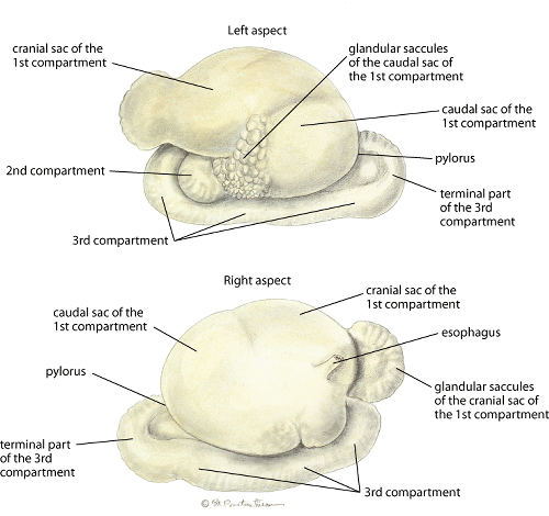
The oral cavity and esophagus of SACs are unremarkable. The stomach has three distinct compartments (C-1, 2, and 3) that do not correlate directly with the four chambers of the ruminant stomach. Although not classed as ruminants, they do eructate, regurgitate, and remasticate and would be considered as foregut fermenters in C-1 and 2. Only the distal fifth of C-3 is analogous to the acid-secreting monogastric stomach. The spiral colon is generally a flat, single spiral prone to blockage when the centripetal loop turns to become centrifugal.
Stomach, llama
Megaesophagus:
Moderate to severe dilatation of the esophagus is relatively common in llamas and alpacas, especially after instances of choking. Signs include chronic weight loss frequently associated with postprandial regurgitation or “frothing” of food. There is no identified age or sex predilection, and no consistent cause has been established. A suspected case of megaesophagus should be confirmed with barium contrast radiography. No treatments (surgical or changes in feeding practices) have been consistently successful. The longterm prognosis is fair to poor, with some animals maintaining condition for an extended period and others continuing to lose weight.
Stomach Atony:
Gastric atony is an occasional problem of unknown cause. Signs include decreased or complete cessation of food consumption, loss of body condition, and depression. Other GI problems, including diarrhea, may be present. Supportive therapy, including fluids, is frequently helpful. Lack of food for 3–5 days also usually causes the death of bacteria and protozoa in C-1 and C-2. Transfaunation (0.5–1 L) of camelid C-1 or strained rumen contents (sheep or cow), administered by gavage, frequently results in a dramatic improvement in appetite and reestablishment of appropriate flora.
Ulcers:
Partial and complete thickness erosions of the acid-secreting distal portion of C-3 and most proximal portion of the duodenum occur. Signs may include decreased food consumption, intermittent to severe colic, and depression. Although the cause has not been clearly established, stress appears to be a significant component, with problems often developing 3–5 days after change of environment affecting social structure, serious injuries, and illnesses.
No reliable premortem diagnostic procedures are available; treatment is usually based on history and clinical signs. Theoretically beneficial oral medications have not proved effective. Parenteral administration of omeprazole reduces acid production. Stress reduction, including clinical housing with a cohort animal, parenteral antibiotics, and supportive therapy, are helpful.
Hepatic Disease:
The visceral surface of the liver normally has multiple fissures, whereas the parietal surface is smooth and lobation is indistinct. There is no gallbladder. SACs appear to be particularly susceptible to Fasciola hepatica, with fecal shedding beginning 10–12 wk after infection. Clinical signs can include ill thrift, diminished growth, and acute death. Icterus is rarely seen. Increased serum bile acids (>25 μmol/L) and enzyme concentrations (normal, alkaline phosphatase 15–121 IU/L and AST 66–235 IU/L) are also diagnostically useful.
Hepatic lipidosis is a relatively common problem in SACs. Clinical signs associated with liver failure in other species are frequently seen, although acute death without prior indication of pending problems also has been reported. The cause is not clearly established, but stress and/or abrupt decrease or change in food consumption appear to play a role. Treatment is symptomatic. Mortality in untreated animals is frequently high.
Small- and Large-intestinal Diseases:
Diarrhea is relatively uncommon in llamas and alpacas. Shortly after birth, SAC crias may experience a mild diarrhea due to abundant dam milk production, essentially a substrate purge. The primary recognized infectious causes of diarrhea in neonates include rotavirus, coronavirus, cryptosporidia, and enteropathogenic strains of Escherichia coli. Some crias also have a transitory diarrhea 2–3 wk after birth, at about the time they experience new food matter. At this same time, some crias develop colic signs due to blockage in the spiral colon. Diarrhea in older neonates is more likely associated with Eimeria spp infection, especially associated with the stress of weaning. Identified causes of diarrhea in older animals include Yersinia pseudotuberculosis, Salmonella spp, Giardia spp, and Cryptosporidium parvum. Treatment options are the same as for other species (ie, fluid and electrolyte replacement and appropriate antibacterials).
Diarrhea in adult SACs is relatively rare but often accompanies a change of feed. Serious conditions characterized by diarrhea include eosinophilic enteritis, infection with Eimeria macusaniensis or Mycobacterium paratuberculosis, or severe nematode parasitism. Compared with that in cattle with Johne's disease, the clinical course in SACs tends to be short and fatal. When diagnosed by fecal examination, E macusaniensis must be promptly treated, because infection can cause marked debilitation. Although variable, current therapy recommendations include oral ponazuril followed by parenteral sulfadimethoxine.
Lymphosarcoma is the only neoplasia found with significant frequency in SACs. It can occur as either a juvenile lymphoma or a primitive malignant round cell tumor. Clinical signs and course vary depending on organ involvement.
Respiratory Diseases
Auscultation of llamas and alpacas is difficult and frequently unrewarding. Little air movement is heard under normal conditions, and identification of areas of infection, congestion, or consolidation is typically difficult. Lateral radiographs may be required for diagnosis of pneumonia. Bacterial infections of the lung are relatively rare, with Streptococcus and Corynebacterium spp being the most common isolates.
Chronic obstructive pulmonary disease appears to be increasing in frequency. Animals allowed to live to their expected life span is a factor, but feeding practices can contribute to onset as well as exacerbation of clinical signs—coughing, shortness of breath, and expiratory dyspnea. Therapy includes changing the feeding regimen to reduce dust, molds, and pollen. Bronchodilators and steroids may be helpful but remain unproved.
Skin Diseases
Unique features of normal camelid skin histologically include a marked vascularity and significant presence of eosinophils. Several skin conditions are shared with sheep and goats, including ringworm, contagious ecthyma, dermatophilosis, and occasionally pizzle rot.
Shearing Injury and Sunburn:
Complications associated with shearing are common. Lacerations may occur where there are loose folds of skin, eg, near the axilla. These often heal uneventfully with or without suturing. Hot shears may cause burns that lead to thick scabs, usually on the dorsum of the back, that may resemble “wool rot.” A history of shearing by a novice often helps confirm the diagnosis. Antibiotic ointment is generally beneficial for these iatrogenic lesions.
Sunburn also can occur after shearing, especially in light-skinned animals. If found in the acute stage, protection from further exposure and application of aloe vera lotion have proved useful. Later appearance of sunburned sites varies from mild peeling to ulcers.
Ulcerative Pododermatitis:
Llamas and alpacas kept in moist conditions develop “immersion foot,” characterized by footpad blistering and sloughing, with variations depending on infection by anaerobic bacteria. Debridement, antiseptics, and foot protection may be required for prolonged periods to facilitate resolution. Treatment with penicillin is always indicated unless unique bacterial isolates are involved. These cases require a relatively long healing period.
Mange, Lice, and Ticks:
All four genera of mange mites (ie, Sarcoptes, Psoroptes, Chorioptes, and Demodex) have been diagnosed in camelids. Alopecia, hyperkeratosis, and scaling accompanied by pruritus tend to characterize all species. The clinical signs may resemble those of zinc deficiency. Deep skin scrapings or biopsies are ideal to make a definitive diagnosis. Although various options for therapy exist, most mange cases will respond to routine parenteral doses of ivermectin repeated every 10–14 days. Oral therapy does not appear to be as effective. Chorioptes infestation may require higher doses repeated every 14–21 days and local therapy. Refractory Sarcoptes cases involving the lower legs have benefited from the same topical treatment.
With louse infestation, it is important to determine whether pediculosis is due to biting (Damalinia breviceps) or sucking (Microthoracius cameli) lice. This can be accomplished with the aid of a hand lens or microscope. Use of transparent tape to retrieve lice for diagnosis from within the depths of wool can be attempted. Sucking lice can be treated with injectable ivermectin as per routine mange therapy. However, biting lice are not affected by parenteral ivermectin. Topical application of synthetic pyrethrin preparations has been effective, but critical doses for these species have not been established. Preventive measures for lice and mange include routine treatment of new herd additions as well as animals visiting and returning for breeding purposes or from shows. Ticks have caused tick paralysis as is seen in other species. In addition, ticks gaining access to ears have caused inner ear afflictions resulting in Horner syndrome as well as encephalitic death.
Copper Deficiency:
Copper deficiency is characterized by depigmentation of fiber with a wiry or steely texture. Juveniles grow poorly and are predisposed to infections. Confirmation of deficiency is best based on comparison of liver copper levels with species normals. Therapy requires dietary supplementation. However, excessive supplementation will cause copper toxicity, which has been diagnosed more commonly than deficiency.
Dorsal Nasal Alopecia (Dark Nose Syndrome):
The most common clinical sign of dorsal nasal alopecia is usually alopecia over the bridge of the nose. The skin is normal or variably scaly, hyperpigmented, and thickened. Dark-haired animals are predisposed, presumably because insects prefer the warmer surface of a dark background. In some animals, the condition may be secondary to rubbing the nose; in others, it may be a fly bite exacerbation. Systemic or topical steroids produce some transient response, but steroids may cause abortion in camelids. In northern climates, the condition tends to spontaneously improve during winter months. Alopecia of the ears has also been seen, particularly in black alpacas.
Idiopathic Hyperkeratosis (Zinc-responsive Dermatosis):
Onset of idiopathic hyperkeratosis is possible at any age. The lesions appear as nonpruritic papules with a tightly adherent crust. Papules progress to plaques and then large areas of thickening and crusting. Lesions are most common in the less densely haired areas of the perineum, ventral abdomen, inguinal region, medial thighs, axilla, and medial forearms, but the face may also be involved. The signs may wax and wane. Diagnosis is by skin biopsy. Treatment is 1 g zinc sulfate or 2–4 g zinc methionine per day. Calcium supplementation should be minimized and alfalfa hay discontinued. Affected animals may be zinc responsive but not deficient.
Idiopathic Nasal/Perioral Hyperkeratotic Dermatosis (Munge):
Most animals with munge are 6 mo to 2 yr old at onset. Variable degrees of hyperkeratosis (heavy, adherent crusts) in paranasal and perioral regions are seen. Less commonly, the bridge of the nose and periocular and periaural regions are affected. Inflammatory lesions may wax and wane. Differential diagnoses include viral contagious pustular dermatitis, dermatophilosis, dermatophytosis, bacterial dermatitis, and autoimmune/immune-mediated disease. Treatment is directed at resolving secondary bacterial infections (ie, daily 10% povidone iodine scrubs plus application of 7% tincture of iodine). If lesions do not respond to antibiotics, adding topical glucocorticoid preparations or intralesional triamcinolone acetonide (2 mg/mL) may be beneficial. Some animals do not respond to any of the described therapies, including those with juvenile llama deficiency syndrome, which has been shown to affect both llamas and alpacas; in these cases, an evaluation of the immune response is indicated.
Resources In This Article
- Llamas and Alpacas
- Overview of Llamas and Alpacas
- Management of Llamas and Alpacas
- Reproduction of Llamas and Alpacas
- Herd Health of Llamas and Alpacas
- Diseases of Llamas and Alpacas


