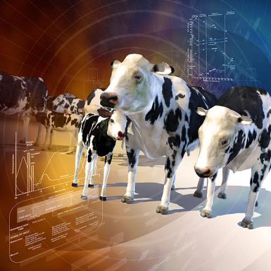
Manipulation of the reproductive system in breeding management programs allows the rapid and dramatic alteration of the conformation and productivity of domestic animals. The reproductive system is incredibly complex in both its anatomy and physiology, and all aspects must be considered when resolving reproductive problems.
Reproductive System

-
Reproductive System Introduction
-
Congenital and Inherited Anomalies of the Reproductive System
-
-
Abortion in Large Animals
- Overview of Abortion in Large Animals
-
- Noninfectious Causes
-
- Neosporosis:
- Bovine Viral Diarrhea (BVD):
- Infectious Bovine Rhinotracheitis (IBR, Bovine Herpesvirus 1):
- Leptospirosis:
- Brucellosis:
- Mycotic Abortion:
- Trueperella pyogenes :
- Trichomoniasis:
- Campylobacteriosis:
- Listeriosis:
- Chlamydiosis:
- Ureaplasma diversum Infection:
- Epizootic Bovine Abortion (Foothill Abortion):
- Bluetongue:
- Other Causes of Abortion:
-
-
-
- Noninfectious Causes
-
- Porcine Reproductive and Respiratory Syndrome (PRRS):
- Porcine Parvovirus:
- Pseudorabies (Aujeszky Disease, Porcine Herpesvirus 1 Infection):
- Japanese B Encephalitis Virus Infection:
- Classical Swine Fever (Hog Cholera):
- Porcine Circovirus Infection:
- Leptospirosis:
- Brucellosis:
- Other Infectious Causes of Abortion:
-
-
-
Bovine Genital Campylobacteriosis
-
Brucellosis in Large Animals
-
Contagious Agalactia
-
Cystic Ovary Disease
-
Equine Coital Exanthema
-
Mastitis in Large Animals
-
Metritis in Large Animals
-
Posthitis and Vulvitis in Sheep and Goats
-
Postpartum Dysgalactia Syndrome and Mastitis in Sows
-
Prolonged Gestation in Cattle and Sheep
-
Pseudopregnancy in Goats
-
Retained Fetal Membranes in Large Animals
-
Seminal Vesiculitis in Bulls
-
Trichomoniasis
-
Udder Diseases
-
Uterine Prolapse and Eversion
-
Vaginal and Cervical Prolapse
-
Vulvitis and Vaginitis in Large Animals
-
Reproductive Diseases of the Female Small Animal
-
Overview of Reproductive Diseases of the Female Small Animal
- Dystocia in Small Animals
- False Pregnancy in Small Animals
- Follicular Cysts in Small Animals
- Mammary Hypertrophy in Cats
- Mastitis in Small Animals
- Metritis in Small Animals
- Ovarian Remnant Syndrome in Small Animals
-
- Subinvolution of Placental Sites in Small Animals
- Vaginal Hyperplasia in Small Animals
- Vaginitis in Small Animals
-
-
Reproductive Diseases of the Male Small Animal
-
Brucellosis in Dogs
-
Mammary Tumors
-
Prostatic Diseases
-
Canine Transmissible Venereal Tumor
Reproductive System Sections (A-Z)
Abortion in Large Animals
Abortion is the termination of pregnancy after organogenesis is complete but before the expelled fetus can survive. If pregnancy ends before organogenesis, it is called early embryonic death. A dead, full-term fetus is a stillbirth (its lungs are not inflated). Many etiologies of abortion also cause stillbirths, mummification, and weak or deformed neonates.
Bovine Genital Campylobacteriosis
Brucellosis in Dogs
Although dogs occasionally become infected with Brucella abortus, B suis, or B melitensis, these sporadic occurrences typically are closely associated with exposure to infected domestic livestock (see Brucellosis in Large Animals).
Brucellosis in Large Animals
Brucellosis is caused by bacteria of the genus Brucella and is characterized by abortion, retained placenta, and to a lesser extent, orchitis and infection of the accessory sex glands in males. The disease is prevalent in most countries of the world. It primarily affects cattle, buffalo, bison, pigs, sheep, goats, dogs (see Brucellosis in Dogs), elk, and occasionally horses. The disease in people, sometimes referred to as undulant fever, is a serious public health problem, especially when caused by B melitensis.
Canine Transmissible Venereal Tumor
Canine transmissible venereal tumors (TVTs) are cauliflower-like, pedunculated, nodular, papillary, or multilobulated in appearance. They range in size from a small nodule (5 mm) to a large mass (>10 cm) that is firm, though friable. The surface is often ulcerated and inflamed and bleeds easily. TVTs may be solitary or multiple and are almost always located on the genitalia. The tumor is transplanted from site to site and from dog to dog by direct contact with the mass. They may be transplanted to adjacent skin and oral, nasal, or conjunctival mucosae. The tumor may arise deep within the preputial, vaginal, or nasal cavity and be difficult to see during cursory examination. This may lead to misdiagnosis if bleeding is incorrectly assumed to be hematuria or epistaxis from other causes. Initially, TVTs grow rapidly, and more rapidly in neonatal and immunosuppressed dogs. Metastasis is uncommon (5%) and can occur without a primary genital tumor present. When metastasis occurs, it is usually to the regional lymph nodes, but kidney, spleen, eye, brain, pituitary, skin and subcutis, mesenteric lymph nodes, and peritoneum may also be sites.
Congenital and Inherited Anomalies of the Reproductive System
Sex determination of the gonads is important for development of the sex phenotype (internal and external genitalia, secondary characteristics) and sexual behavior. A sex chromosome genotype of XY leads to the development of testes due to the sex-determining region of the Y chromosome (SRY) gene. The SRY gene induces downstream factors such as SRY-box containing gene 9 (SOX9), anti-Müllerian hormone, and glial cell line–derived neurotrophic factor in Sertoli cells.
Contagious Agalactia
First recognized in Italy more than 200 years ago, contagious agalactia is primarily a disease of dairy sheep and goats and is characterized by an interstitial mastitis leading to a loss of milk production, arthritis, and infectious keratoconjunctivitis. It is more often seen on farms practicing traditional husbandry. Contagious agalactia is principally caused by the wall-less bacterium Mycoplasma agalactiae, but in recent years, M mycoides capri (Mmc; formerly known as LC), and, to a lesser extent, M capricolum capricolum (Mcc) and M putrefaciens have also been isolated from goats with mastitis, arthritis, and occasionally, respiratory disease. The clinical signs of these infections are sufficiently similar to those of contagious agalactia for the OIE to include them as causes of this listed disease.
Cystic Ovary Disease
Among domestic animals, cystic ovary disease (COD) is most common in cattle, particularly the dairy breeds, but it occurs sporadically in dogs (see Follicular Cysts in Small Animals), cats, pigs, and perhaps mares.
Equine Coital Exanthema
Mammary Tumors
The frequency of mammary neoplasia in different species varies tremendously. The dog is by far the most frequently affected domestic species, with a prevalence ~3 times that in women; ~50% of all tumors in the bitch are mammary tumors. Mammary tumors are rare in cows, mares, goats, ewes, and sows. There are differences in both biologic behavior and histology of mammary tumors in dogs and cats. Approximately 45% of mammary tumors are malignant in dogs, whereas ~90% are malignant in cats, and dogs have a much higher number of complex and mixed tumors than do cats.
Mastitis in Large Animals
Mastitis, or inflammation of the mammary gland, is predominantly due to the effects of infection by bacterial pathogens, although mycotic or algal microbes play a role in some cases. Pathologic changes to milk-secreting epithelial cells from the inflammatory process often bring about a decrease in functional capacity. Depending on the pathogen, functional losses may continue into further lactations, which may reduce productivity and potential weight gain for suckling offspring. Although most infections result in relatively mild clinical or subclinical local inflammation, more severe cases can lead to agalactia or even profound systemic involvement, resulting in death. Mastitis has been reported in almost all domestic mammals and has a worldwide geographic distribution. Climatic conditions, seasonal variation, bedding, housing density of livestock populations, and husbandry practices may affect the incidence and etiology. However, it is of greatest frequency and economic importance in species that primarily function as producers of milk for dairy products, particularly dairy cattle. (Also see Udder Diseases.)
Metritis in Large Animals
In all species, acute puerperal metritis occurs within the first 10–14 days postpartum. It results from contamination of the reproductive tract at parturition and often, but not invariably, follows complicated parturition. Important causative organisms in cattle include Escherichia coli and Trueperella (Arcanobacterium) pyogenes, but culture-independent studies have demonstrated the dominant role of gram-negative anaerobic bacteria such as Prevotella melaninogenica and Fusobacterium necrophorum. The condition is usually acute in onset. Affected cows, mares, ewes, does, or sows are depressed, febrile, and inappetent. A fetid, watery uterine discharge is characteristic of the condition in cows but may not be conspicuous in other species. Milk production is diminished, and nursing young may show signs of food deprivation.
Posthitis and Vulvitis in Sheep and Goats
Two common and distinct forms of posthitis and vulvitis are recognized in small ruminants. The first, referred to as enzootic posthitis and vulvitis, is associated with high-protein diets, infection with Corynebacterium renale or other urease-producing organisms, locally high concentrations of ammonia, and severe posthitis. The second is referred to as necrotic or ulcerative balanoposthitis and vulvitis. Its cause is unclear, but Mycoplasma mycoides mycoides is implicated, as are other Mycoplasma spp organisms of the Histophilus/Haemophilus group, and potentially viruses, such as caprine herpesvirus 1.
Postpartum Dysgalactia Syndrome and Mastitis in Sows
Numerous etiologies or pathophysiologies can be involved in this syndrome, which is reflected by the use of several different names—mastitis-metritis-agalactia (MMA) complex, agalactia syndrome, dysgalactia syndrome, mammary edema, periparturient hypogalactia syndrome, agalactia toxemia, and puerperal mastitis. However, these names are not synonymous and have often been misused. The syndrome can currently be classified according to the number of mammary glands affected, ie, uniglandular or multiglandular mastitis (including postpartum dysgalactia syndrome [PPDS], MMA complex). (Also see Mastitis in Large Animals.)
Prolonged Gestation in Cattle and Sheep
Parturition is induced by the fetus in both cattle and sheep. It is initiated by rising cortisol levels in the fetus that provoke a cascade of endocrine activity in the dam. Fetal cortisol increases as a result of increased adrenocorticotropic hormone (ACTH) production by the maturing fetal pituitary caused by fetal stressors such as hypoxia and hypercapnia. Gestation length is unique to each fetus, but approximate gestation lengths can be ascribed to each species (see Table: Approximate Gestation Periods).
Prostatic Diseases
Disease of the prostate gland is relatively common in intact dogs but less common in other domestic animal species. Benign prostatic hyperplasia is by far the most common disease of the prostate in intact male dogs. Bacterial prostatitis (acute or chronic), prostatic abscesses, prostatic and paraprostatic cysts, and prostatic adenocarcinoma are seen much less frequently and can be seen in castrated males. Depending on the disorder, clinical signs may include tenesmus during defecation, intermittent hematuria, recurrent urinary tract infections, and caudal abdominal discomfort. However, many intact males with benign prostatic hyperplasia (with or without chronic prostatitis) are asymptomatic or present with signs of hemospermia and/or infertility only. Additional nonspecific signs, such as fever, malaise, anorexia, severe stiffness, and caudal abdominal pain, can be seen with acute bacterial infections, abscesses, and neoplasia. Prostatic adenocarcinoma with bony involvement of the pelvis and lumbar vertebrae may cause hindlimb gait abnormalities. Less commonly, prostatic diseases may cause urinary incontinence. Prostatic adenocarcinoma may cause complete urethral obstruction.
Pseudopregnancy in Goats
Reproductive Diseases of the Female Small Animal
Reproductive Diseases of the Male Small Animal
Reproductive System Introduction
The reproductive system provides the mechanism for the recombination of genetic material that allows for change and adaptation. Manipulation of this system in breeding management programs allows the rapid and dramatic alteration of the conformation and productivity of domestic animals. Theriogenology is the veterinary clinical specialty that deals with reproduction. The reproductive system is incredibly complex in both its anatomy and physiology, and all aspects must be considered when resolving reproductive problems. The differences in the reproductive system between the sexes and among species are extensive. Both sexes have primary sex organs and primary regulatory centers. Gonads and function-adapted, tubular genital organs constitute primary sex organs in both sexes. The pituitary gland and the hypothalamus are the primary regulatory centers; thus, the regulatory function is, in part, neuroendocrine in nature. In pregnant females, the fetoplacental unit has a significant role in maintaining and terminating pregnancy.
Retained Fetal Membranes in Large Animals
Retention of fetal membranes, or retained placenta, usually is defined as failure to expel fetal membranes within 24 hr after parturition. Normally, expulsion occurs within 3–8 hr after calf delivery. The incidence in healthy dairy cows is 5%–15%, whereas the incidence in beef cows is lower. The incidence is increased by abortion (particularly with brucellosis or mycotic abortion), dystocia, twin birth, stillbirth, hypocalcemia, high environmental temperature, advancing age of the cow, premature birth or induction of parturition, placentitis, and nutritional disturbances. Cows with retained fetal membranes are at increased risk of metritis, displaced abomasum, and mastitis.
Seminal Vesiculitis in Bulls
The seminal vesicles are paired accessory sex glands located on the floor of the pelvis, lateral to the ampullae, and dorsal to the neck of the urinary bladder. The vesicles secrete a clear fluid that adds volume, nutrients, and buffers to semen. The term “seminal” vesicle is a misnomer, because the vesicles are not a reservoir for spermatozoa.
Trichomoniasis
Udder Diseases
Also see Mastitis in Large Animals and Pseudocowpox.
Uterine Prolapse and Eversion
Prolapse of the uterus may occur in any species; however, it is most common in dairy and beef cows and ewes and less frequent in sows. It is rare in mares, bitches, queens, and rabbits. Invagination of the tip of the uterus, excessive traction to relieve dystocia or retained fetal membranes, uterine atony, hypocalcemia, and lack of exercise have all been incriminated as contributory causes.
Vaginal and Cervical Prolapse
Eversion and prolapse of the vagina, with or without prolapse of the cervix, occurs most commonly in cattle and sheep. A form of vaginal prolapse, different in pathogenesis, also occurs in dogs (see Vaginal Hyperplasia in Small Animals). In cattle and sheep, the condition is usually seen in mature females in the last trimester of pregnancy. Predisposing factors include increased intra-abdominal pressure associated with increased size of the pregnant uterus, intra-abdominal fat, or rumen distention superimposed upon relaxation and softening of the pelvic girdle and associated soft-tissue structures in the pelvic canal and perineum mediated by increased circulating concentrations of estrogens and relaxin during late pregnancy. Intra-abdominal pressure is increased in recumbent animals. Added to this, sheep tend to face uphill when lying down, so gravity contributes to vaginal eversion and prolapse. Docking the tails of lambs may damage structures that support the pelvic girdle (eg, coccygeus muscle) and predispose to vaginal prolapse if the tail is docked too short. The tail should be removed at the level of the ventral skin fold, leaving two or three coccygeal vertebrae intact.
Vulvitis and Vaginitis in Large Animals
Contusion and hematoma of the vagina are noted infrequently after parturition in all species but particularly in mares and sows. Occasionally, vaginal hematomas in sows may rupture and cause serious (or fatal) hemorrhage that can be controlled by ligation of the labial branch of the internal pudendal artery. Necrotic vaginitis, vestibulitis, and vulvitis may follow dystocia in all species. Onset of signs, consisting of arched back, elevated tail, anorexia, dysuria, straining, vulvar and perivulvar swelling, and possibly a fetid, serous discharge, begins within 1–4 days of parturition and may persist for 2–4 wk. In most cases, only gentle and conservative treatment is needed. Prophylactic antibiotic treatment is wise, because clostridial or other organisms may proliferate in the damaged tissue and cause tetanus (see Tetanus), blackleg (see Blackleg), or other forms of clostridial myositis. Possible consequences of necrotic vaginitis include permanent stricture of the vagina, transvaginal adhesions, or perivaginal abscessation.
Also of Interest
Test your knowledge
Mastitis in dairy cows is most commonly caused by a bacterial infection. The sources of these infections are typically environmental or contagious. Which of the following organisms is most likely to be spread between cows via aerosol transmission?


