


Follicular Cystic Ovary Disease in Cattle
(Cystic follicles, “Bulling”)
- Cystic Ovary Disease
- Overview of Cystic Ovary Disease in Large Animals
- Follicular Cystic Ovary Disease in Cattle
- Luteal Cystic Ovary Disease in Cattle
- Cystic Ovary Disease as a Herd Problem
- Cystic Ovary Disease in Mares
Follicular cystic ovary disease may be defined on a number of levels. Essentially, all signs relate to the disruption of the normal endocrine events of the estrous cycle, through the failure of ovulatory events. Key features include the presence of a thin-walled, fluid-filled structure >25 mm diameter and present for >7 days in the absence of a CL on ultrasonographic imaging of the ovary. Along with occasional behavioral signs of nymphomania, such as short inter-estrus intervals and excessive heat behavior, are endocrine changes, including a suboptimal luteinizing hormone (LH) surge and persistently increased estradiol levels.
Etiology and Pathogenesis:
A hereditary predisposition has been implicated in dairy cattle. Cystic ovary disease or syndrome (COD) is commonly considered to be associated with negative energy balance and stress factors in dairy cows that are high milk producers. Genetic predisposition to partition glucose to the udder to prioritize lactose synthesis and milk production is characterized by insulin resistance in Holstein cattle. IGF-1 is a cytokine associated with metabolic influences on reproduction and hence COD. Incidence increases with age. Most cases occur within 3–8 wk of parturition at the first attempted postpartum ovulation, coinciding with peak daily milk production and rapidly decreasing body condition. The reported herd incidence is 5%–25% per lactation, or higher in some problem herds.
During normal proestrus, regression of the CL coincides with development of a selected follicle, while the growth of any additional follicles is inhibited. In animals developing COD, ovulation fails to occur and the dominant follicle continues to enlarge. An important component of the etiology is the failure of positive feedback of follicular estrogen on the hypothalamus via estrogen receptor α to release sufficient GnRH during estrus to trigger an LH surge. The end result is a failure of ovulation at the time of estrus. Moreover, other follicles may grow and form multiple cysts either bilaterally or unilaterally.
Grossly, follicular cysts resemble enlarged follicles, generally defined as varying in size from 25 mm to 50–60 mm in diameter. The size and form of an affected ovary depends on the number and size of cysts present. The cystic ovary is capable, at least initially, of steroidogenesis, and its products vary from estrogens to progesterone to androgens. The actions of the various hormones produced or the absence of the stabilizing action of high progesterone from the normal CL during ~75% of the estrous cycle (or both) are responsible for the changes seen in the genital tract, body conformation, and general behavior.
Clinical Findings:
Behavioral aberrations range from frequent, intermittent estrus with exaggerated monosexual drive to bull-like behavior, including mounting, pawing the ground, and bellowing. This behavior may be accompanied by masculinization of the head and neck. Relaxation of the vulva, perineum, and the large pelvic ligaments, which causes the tail head to be elevated, can occur in chronic cases. Some affected cows show these signs, but others may be sexually quiescent; anestrous or subestrous cows are a common presentation.
The affected ovaries generally are enlarged and rounded, but their size varies, depending on the number and size of cysts. Their surface is smooth, elevated, and blister-like. Cysts frequently are multiple and may approach 4–6 cm in diameter. Under the influence of hormones produced by the cystic ovary or the lack of hormones (especially progesterone) normally present during estrous cycles, the uterus undergoes palpable changes, which in turn vary with the duration of the cystic condition. Thus, during the first week, the uterine wall is thickened and edematous as an extension of the preceding estrus. Toward the end of the first week, the uterine wall develops a sponge-like consistency. In chronic cases, atony and atrophy of the uterine wall are common. Occasionally, the uterine horns become markedly shortened. Some degree of mucoid to mucopurulent vaginal discharge is common. Hydrometra, a fluid-filled, extremely thin-walled uterus, is seen occasionally.
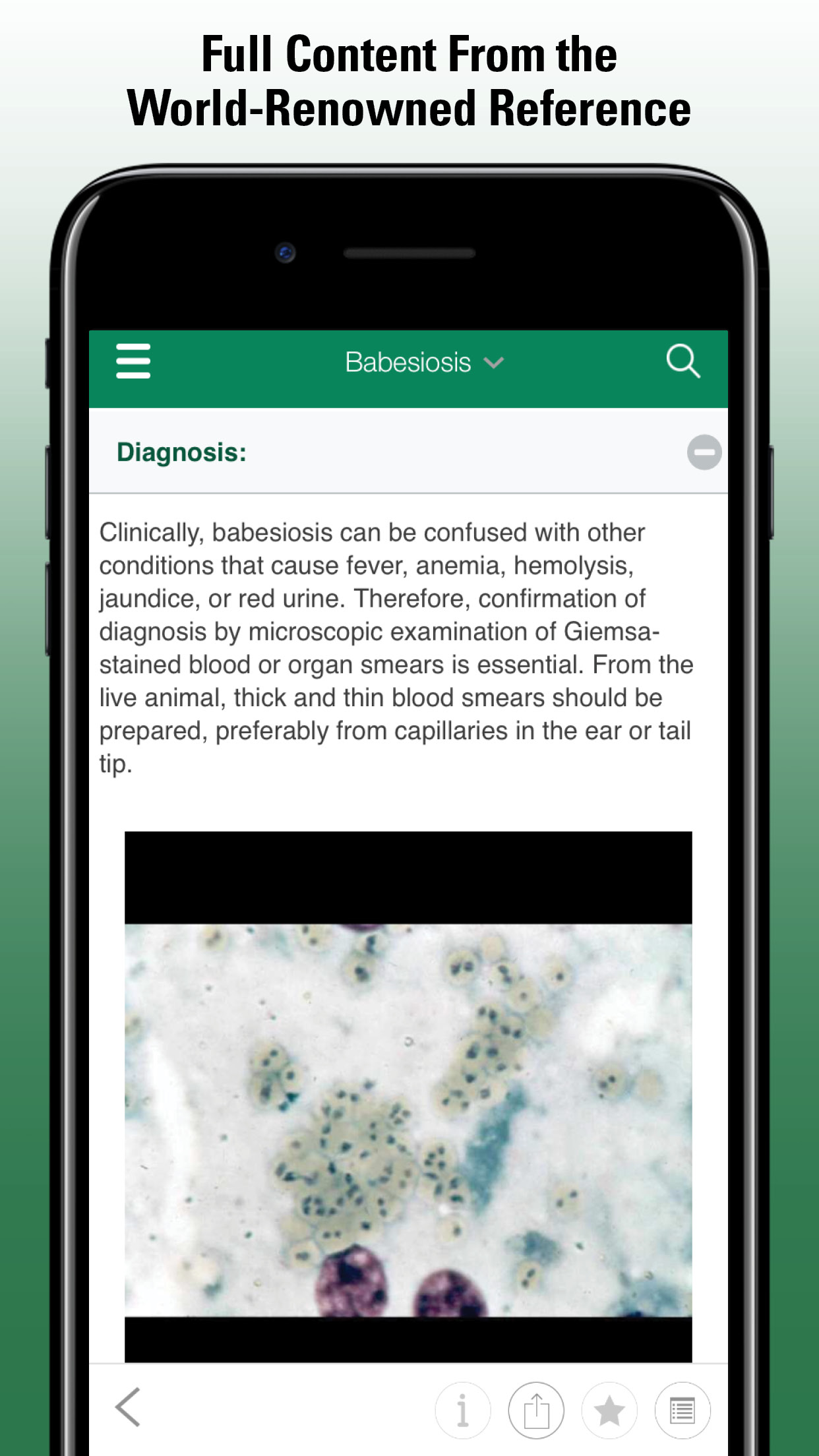
Diagnosis:
Palpation of the uterus may help differentiate a follicular cyst from a dominant preovulatory follicle; the estrous cow has a coiled, extremely turgid uterus and a follicle. As noted earlier, cystic cows fail to ovulate a preovulatory follicle after undergoing CL regression and, on examination of the reproductive tract, they present with a large follicle, absence of a CL, and absence of a turgid uterus. Ultrasound technology per rectum can be used to differentiate cysts from corpora lutea and may help diagnose cyst type (ie, follicular cyst wall diameter has been described as <3 mm vs luteal cyst wall diameter >3 mm). Larger, multiple cysts are easily identified by rectal palpation. History, conformation, and uterine changes, when present, provide supplemental diagnostic evidence.
Treatment:
COD may be frustratingly unresponsive to therapy. Some cysts respond readily to an LH-type hormonal treatment. In the past, human chorionic gonadotropin (hCG) was commonly used. It is most effective at 10,000 USP units IM, although success with lower doses given IM or IV has also been reported.
Hormone therapy with GnRH may be effective at 100 mcg and less antigenic than hCG. To hasten the onset of the first estrus after treatment, prostaglandin (PG) F2α products can be given 7 days after hCG or GnRH. Ovulation synchronization protocols combine GnRH and PGF2α to control follicular dynamics, luteolysis, and ovulation. They allow for fixed timed artificial insemination (TAI) of cattle without the need for estrus detection and have been successfully used to treat cows with cystic ovaries. This protocol consists of giving GnRH, then prostaglandin 7 days later, then a second administration of GnRH 48 hr later, and finally TAI 0–24 hr later. Breeding on the first estrus may reduce recurrence by establishing pregnancy as soon as possible. However, COD recurrence is a risk.
Manual rupture is used, but the potential danger of traumatizing the ovary and causing hemorrhage with subsequent local adhesions should not be overlooked.
Prognosis:
After therapy with an LH-type hormone, a normal, fertile estrus can be expected in 15–30 days. With GnRH therapy, 25% of cases required a second treatment, and 5% required a third. One-third of the cases treated for the third time did not respond. Spontaneous recovery is possible and is most common in cases arising during the first 50 days after calving. Likewise, successful treatment encourages perpetuation of the disease in the herd if the offspring are used for breeding. Although COD in cattle clearly has a genetic component, it is unlikely that a single farm using artificial insemination can significantly influence the incidence. In Sweden, progress has been made in reducing the condition through culling and selection procedures for bulls used in artificial insemination, but affected cows are often still treated.
- Cystic Ovary Disease
- Overview of Cystic Ovary Disease in Large Animals
- Follicular Cystic Ovary Disease in Cattle
- Luteal Cystic Ovary Disease in Cattle
- Cystic Ovary Disease as a Herd Problem
- Cystic Ovary Disease in Mares
Also of Interest
Test your knowledge
Mastitis in dairy cows is most commonly caused by a bacterial infection. The sources of these infections are typically environmental or contagious. Which of the following organisms is most likely to be spread between cows via aerosol transmission?