Laminitis in Horses
(Founder)
- Lameness in Horses
- Overview of Lameness in Horses
- The Lameness Examination in Horses
- Imaging Techniques in Equine Lameness
- Arthroscopy in Equine Lameness
- Regional Anesthesia in Equine Lameness
- Disorders of the Foot in Horses
- Osseous Cyst-like Lesions in the Distal Phalanx in Horses
- Bruised Sole and Corns in Horses
- Canker in Horses
- Fracture of Navicular Bone in Horses
- Fracture of Distal Phalanx in Horses
- Keratoma in Horses
- Laminitis in Horses
- Navicular Disease in Horses
- Pedal Osteitis in Horses
- Puncture Wounds of the Foot in Horses
- Pyramidal Disease in Horses
- Quittor in Horses
- Quarter Crack in Horses
- Scratches in Horses
- White Line Disease in Horses
- Sheared Heels in Horses
- Sidebone in Horses
- Thrush in Horses
- Disorders of the Pastern and Fetlock
- Fractures of the First and Second Phalanx in Horses
- Fractures of the Proximal Sesamoid Bones in Horses
- Osteoarthritis of the Proximal Interphalangeal Joint in Horses
- Palmar/Plantar Metacarpal/Metatarsal Nonadaptive Bone Remodeling in Horses
- Sesamoiditis in Horses
- Chronic Proliferative Synovitis in Horses
- Digital Sheath Tenosynovitis in Horses
- Disorders of the Metacarpus in Horses
- Tendinitis in Horses
- Suspensory Desmitis in Horses
- Inferior Check Desmitis in Horses
- Bucked Shins in Horses
- Exostoses of the Second and Fourth Metacarpal Bones in Horses
- Fractures of the Small Metacarpal (Splint) Bones in Horses
- Fracture of the Third Metacarpal (Cannon) Bone in Horses
- Disorders of the Carpus in Horses
- Fracture of the Carpal Bones in Horses
- Subchondral Bone Disease of the Third Carpal Bone in Horses
- Tearing of the Medial Palmar Intercarpal Ligament in Horses
- Osteoarthritis of the Carpus in Horses
- Distal Radial Exostosis and Osteochondroma of the Distal Radius in Horses
- Carpal Hygroma in Horses
- Rupture of the Common Digital Extensor Tendon in Horses
- Disorders of the Shoulder in Horses
- Developmental Diseases of the Shoulder in Horses
- Fractures of the Shoulder in Horses
- Bicipital Bursitis in Horses
- Infection of the Shoulder in Horses
- Suprascapular Neuropathy in Horses
- Osteoarthritis of the Shoulder in Horses
- Disorders of the Elbow in Horses
- Developmental Orthopedic Disease in the Elbow of Horses
- Fractures of the Elbow in Horses
- Osteoarthritis of the Elbow in Horses
- Collateral Ligament Injury in the Elbow of Horses
- Disorders of the Metatarsus in Horses
- Bucked Shins/Dorsal Cortical Fractures of the Third Metatarsal Bone in Horses
- Exostoses of the Metatarsal Bones in Horses
- Diaphyseal Fracture of the Third Metatarsal Bone in Horses
- Incomplete Longitudinal Fractures of the Plantar Aspect of the Third Metatarsal Bone in Horses
- Focal Bone Reaction and Avulsion Fractures of the Third Metatarsal Bone in Horses
- Fractures of the Second and Fourth Metatarsal Bones in Horses
- Enostosis-like Lesions of the Third Metatarsal Bone in Horses
- Disorders of the Tarsus in Horses
- Failure of Ossification of the Distal Tarsal Bones in Horses
- Osteoarthritis of the Distal Tarsal Joints in Horses
- Osteoarthritis of the Talocalcaneal Joint in Horses
- Osteoarthritis of the Tarsocrural Joint in Horses
- Synovitis/Capsulitis of the Tarsocrural Joint in Horses
- Osteochondrosis of the Tarsocrural Joint in Horses
- Osteitis of the Calcaneus in Horses
- Fractures of the Distal Tarsal Bones in Horses
- Fracture of the Talus in Horses
- Fracture of the Fibular Tarsal Bone (Calcaneus) in Horses
- Fracture of the Lateral Malleolus of the Tibia in Horses
- Tarsal Joint Luxation in Horses
- Desmitis of the Collateral Ligaments of the Tarsus in Horses
- Rupture of the Fibularis (Peroneus) Tertius in Horses
- Stringhalt
- Curb in Horses
- Disorders of the Tarsal Sheath in Horses
- False Thoroughpin in Horses
- Luxation of the Superficial Digital Flexor Tendon from the Tuber Calcanei in Horses
- Gastrocnemius Tendinitis in Horses
- Calcaneal Bursitis in Horses
- Capped Hock
- Disorders of the Stifle in Horses
- Osteochondrosis of the Stifle in Horses
- Subchondral Cystic Lesions in Horses
- Meniscus and Meniscal Ligament Injuries in Horses
- Cranial and Caudal Cruciate Ligament Injuries in Horses
- Collateral Ligament Injuries in Horses
- Intermittent Upward Fixation of the Patella and Delayed Patella Release in Horses
- Fragmentation of the Patella in Horses
- Patellar Luxation in Horses
- Patellar Ligament Injuries in Horses
- Gonitis and Osteoarthritis in Horses
- Chondromalacia of the Femoral Condyles in Horses
- Fractures of the Stifle in Horses
- Disorders of the Hip in Horses
- Luxation of the Coxofemoral Joint in Horses
- Pelvic Fracture in Horses
- Osteoarthritis and other Coxofemoral Joint Diseases in Horses
- Disorders of the Back and Pelvis in Horses
- Spinal Processes and Associated Ligaments in Horses
- Articular Process−Synovial Intervertebral Articulation Complexes in Horses
- Vertebral Bodies and Discs in Horses
- Muscle Strain and Soreness in Horses
- Lumbosacral Junction Abnormalities in Horses
- Sacroiliac Joint Abnormalities in Horses
- Developmental Orthopedic Disease in Horses
- Osteochondrosis in Horses
- Physitis in Horses
- Flexural Deformities in Horses
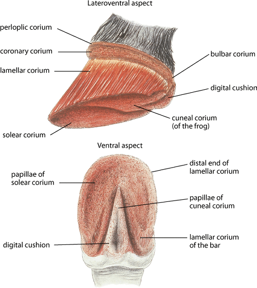
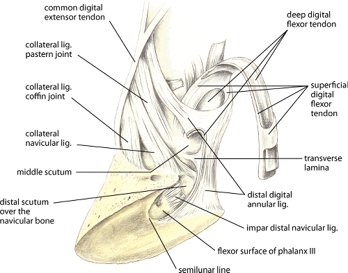
Equine laminitis is a crippling disease in which there is a failure of attachment of the epidermal laminae connected to the hoof wall from the dermal laminae attached to the distal phalanx. Because the laminae are responsible for suspending the distal phalanx within the hoof wall, laminar failure in combination with the downward forces of the weight of the horse and distracting forces such as the tension from the deep digital flexor tendon commonly results in a catastrophic displacement of the distal phalanx, resulting in severe lameness. Laminitis affects all breeds of horses.
Etiology and Pathogenesis:
Three main disease states are thought to be associated with laminitis: 1) diseases associated with sepsis or endotoxemia, 2) endocrinopathic laminitis encompassing both equine metabolic syndrome (including pasture-associated laminitis, see Equine Metabolic Syndrome and pituitary adenoma), and 3) supporting limb laminitis. The pathogenesis of laminitis remains controversial and most likely varies widely between these three primary causes. A fourth, less common cause is ingestion of shavings (sometimes inadvertently used for bedding) from black walnut heartwood; the pathophysiology of this type of laminitis appears similar to that of sepsis-related laminitis. The diseases causing systemic sepsis in sepsis-related laminitis are diseases commonly associated with gram-negative bacterial (or polymicrobial) sepsis and include ingestion of excess carbohydrate (grain overload), acute postparturient metritis (retained fetal membranes), colic (anterior enteritis, large colon volvulus), and enterocolitis. Laminitis secondary to equine metabolic syndrome most commonly occurs in overweight horses and ponies and is commonly exacerbated when grazing lush pastures. It is possible that laminitis occurring from an acute intake of lush pasture may be a combination of sepsis-related laminitis (similar to grain overload) and metabolic syndrome. Supporting limb laminitis can occur any time the horse places excessive weight on one limb for an extended period because of inability to use the other limb (eg, postoperative orthopedic procedures, radial nerve paralysis, or a septic joint or tendon sheath).
The basic cause of laminar failure in laminitis is a failure of attachment of the laminar basal epithelial cells (LBECs) of the epidermal laminae to the underlying dermal laminae. Although this failure was thought to be primarily due to breakdown of the matrix molecules in the basement membrane and dermis (to which the LBECs attach) by matrix metalloproteases, studies have questioned the importance of matrix metalloproteases. It appears that the LBECs may primarily be losing attachment to the underlying dermal laminae due to dysregulation of the hemidesmosomes (the adhesion complexes on the basal side of the LBECs that attach the cells to the underlying matrix molecules of the basement membrane) and possibly the associated cytoskeleton. Inflammatory mediators and enzymes (eg, proinflammatory cytokines, cyclooxygenase-2) are markedly increased in the laminae in the early stages of sepsis-related laminitis and may injure the LBECs or cause cellular dysregulation, leading to loss of attachment. Hypoxia and ischemia due to aberrant vascular flow is also likely to play a role in LBEC dysfunction in sepsis-related laminitis but appears to occur later in the disease process.
The pathophysiology behind laminitis associated with equine metabolic syndrome is not as well researched as sepsis-related laminitis, but work indicates that inflammatory signaling does not play a major role and that dysregulation of the LBECs is likely to result from insulin-related signaling, possibly through growth factor receptors such as IGF-1 receptor. The pathogenesis of supporting limb laminitis is only now being intensively investigated. These studies indicate that the central pathophysiologic factor in supporting limb laminitis is decreased laminar blood flow due to a lack of movement of the supporting limb because of pain in the opposite limb.
After loss of integrity of the laminar attachments, the distal phalanx can undergo three types of displacement depending on the forces placed on the foot and the pattern of laminar injury. Symmetrical distal displacement of the entire phalanx (usually termed “sinking”) occurs when there is circumferential loss of laminar attachments, most commonly seen in severe cases of sepsis but also seen in equine metabolic syndrome. Palmar rotation of the distal margin of the distal phalanx (usually termed “rotation”) is the most common displacement seen, and most likely occurs due to a combination of loss of the dorsal laminar attachments (while maintaining some laminar integrity in the quarters and heels) and tension on the deep digital flexor tendon. Rarely, uniaxial/unilateral distal displacement of the distal phalanx occurs, most commonly to the medial side in the forelimb; this displacement can only be visualized on an anterior-posterior radiograph of the foot. In laminitis related to sepsis and equine metabolic syndrome, the forelimbs are most commonly affected, although the hindlimbs can also be affected in severe cases. In supporting limb laminitis, either a front or rear foot is affected depending on which opposite limb has the weight-bearing problem.
Clinical Findings:
Classically, laminitis is considered acute, subacute, or chronic. Acute laminitis is classically defined as the initial few days of clinical signs of laminitis (usually <3 days) in a horse in which the distal phalanx has not undergone displacement. Subacute laminitis is commonly used to define laminitis in which clinical signs have continued >3 days, but the horse still has no distal phalangeal displacement. Chronic laminitis is classically defined as the case in which distal phalanx displacement has occurred regardless of the duration of the disease. Early in laminitis, the horse is depressed and anorectic and stands reluctantly. Resistance to any exercise is marked, and the normal stance is altered in attempts to relieve the weight borne by the affected feet. If only the forelimbs are affected, the horse will stand with the forelimbs placed far forward (to decrease the weight on the front digits); the hindlimbs also are placed more forward to support more of the weight of the horse. If forced to walk, the horse shows a slow, crouching, short-striding gait. If all four limbs are affected, the animal will appear “camped out,” with the forelimbs placed more forward than usual and the hindlimbs placed more caudally than usual. Each foot, once lifted, is set down as quickly as possible.
In the acute stage of laminitis, the entire hoof wall may be warm. An exaggerated and bounding pulse can be palpated and may be visible in the digital arteries. Pain can cause muscular trembling, and a fairly uniform tenderness can be detected when pressure is applied to the sole (most commonly in the toe region). The pulse rate (60–120/min) and respiratory rate (80–100/min) are commonly increased, primarily because of pain. Lameness is usually moderate to severe at this time. In exceptionally severe cases, for which the prognosis is unfavorable, a blood-stained exudate may seep from the coronary bands. These clinical signs do not always occur in endocrinopathic laminitis because of the insidious nature of the disease process, which can occur over months or years; the first clinical sign in many of these horses is toe bruising due to solar compression by the slowly displacing distal phalanx. Radiographic evidence of displacement of the distal phalanx can be present as early as the third day after the onset of disease in horses with sepsis/endotoxemia. However, an MRI study has shown that, in the acute case, the horse may have normal-appearing distal phalanx on radiographs, despite destruction of the entire dorsal laminar attachment that is visible on MRI.
Subacute cases may exhibit any or all of the above clinical signs but to a lesser degree. Often, there is only a mild change in stance, with reluctance to walk and some increased sensitivity to concussion on the soles of the affected feet. There may be no demonstrable heat in the coronary band or increase in digital pulse. The acute and subacute forms of laminitis tend to recur at varying intervals and may develop into the chronic form.
During and immediately after displacement of the distal phalanx (classically termed chronic laminitis once displacement occurs, regardless of the temporal aspect of the disease course), the horse is usually extremely lame and may spend a great deal of time recumbent. In severe cases, the foot may prolapse through the sole cranial to the frog, or the coronary band may separate; both occurrences gravely affect the prognosis. Longterm cases of chronic laminitis are characterized by changes in the shape of the hoof and usually follow one or more attacks of the acute form. Especially with cases of rotation of the distal phalanx, bands of irregular horn growth (laminitic rings) may be seen in the hoof, close at the toe and diverging at the heel (due to minimal hoof growth from the dorsal coronary band). The hoof itself becomes narrow and elongated, with “dishing” of the dorsal surface of the hoof wall with a steep angle to the hoof wall proximally and a much more horizontally oriented wall distally.
As the displacement of the distal phalanx progresses in longterm cases, the sole becomes flattened or somewhat convex in outline on the ground surface immediately cranial to the apex of the frog. The gait is similar to that already described, and when standing, the body weight is continually shifted from one foot to the other. Radiography commonly reveals rotation and some bony resorption of the dorsal solar margin of the distal phalanx in longer term cases.
Diagnosis:
In acute laminitis, diagnosis is usually straightforward and is based on the history (eg, grain overload) and posture of the horse, increased temperature of the hooves, a hard pulse in the digital arteries, and reluctance to move. Abaxial sesamoid nerve blocks of the forelimb digits in the very lame horse allow assessment of possible involvement of the hindfeet (by walking the animal a few steps) and enable full assessment of the soles of both feet (for solar prolapse, etc). These nerve blocks also make it possible to obtain good quality lateral and anterior-posterior radiographs of the foot without severe duress to the horse. Lidocaine should be used for the nerve block, because it will last only a short time (ie, not long enough for the animal to move excessively and further damage the laminae); applying a temporary pad to the foot not being radiographed (to protect that foot) is also recommended. Gross observation and distinct measurements of the hoof wall and sole thicknesses from the radiographs allow determination of whether distal displacement, rotation, both distal displacement and rotation, or unilateral sinking has occurred. Depending on the radiographic diagnosis of the type of phalangeal displacement present, there is usually time while the nerve block is still effective to apply a temporary type of pad or shoe.
Treatment:
Acute laminitis constitutes a medical emergency, because phalangeal displacement can occur rapidly. Despite prompt therapy, the prognosis is guarded until recovery is complete and it is evident that the hoof architecture is not altered. Most animals should be administered NSAIDs, with flunixin meglumine being the drug of choice if the horse is still systemically ill (ie, enterocolitis). Phenylbutazone is usually used in the early chronic stage when the horse is lame but does not have signs of systemic disease such as sepsis/endotoxemia. Close attention to the potential toxicities of NSAID therapy, particularly with phenylbutazone, is required. Because phenylbutazone accumulates in the tissue (unlike flunixin or most other NSAIDs), it is best to skip a day every 5–7 days to “clear the system” (flunixin can be administered that day). NSAIDs should be used according to label instructions and, if used in combination, the dosage of each drug should be reduced accordingly. Another option for treatment of chronic laminitis in horses at risk of renal or GI complications is the COX-2-selective NSAID firocoxib. Other options for analgesia include detomidine, butorphanol, morphine, or a constant-rate infusion of a “cocktail” of sedatives and analgesics.
For treatment of possible ongoing ischemia, acepromazine is the only drug found to effectively increase digital blood flow in some studies; however, this has been questioned again by a study using laminar microdialysis catheters in which urea clearance (an indicator of blood flow) was not changed in horses administered acepromazine. In a horse at risk of or in the early stages of sepsis-related laminitis, digital hypothermia (cooling of the foot by placing it directly in ice water) has been popularized again by several experimental studies consistently demonstrating efficacy in protection of the structural integrity of the lamellar tissue; in one clinical study of enterocolitis cases in two hospitals, risk of developing laminitis was decreased 10-fold in septic/endotoxemic horses in which continuous digital hypothermia was performed.
During the first 2–3 wk, it is important to remove standard shoes, because shoes place the majority of stress on the hoof wall and therefore the laminae. The feet should be padded with a soft, resilient substance such as a 1- to 2-in. thick piece of closed-cell foam cut to the diameter of the foot. Pads to provide sole support can also be made from different putties available to farriers. Decreasing padding (or beveling the pad) in the region dorsal to the apex of the frog decreases the stress on the dorsal laminae. Styrofoam insulation (2 in. thick) can be used in small equids but usually provides minimal support in larger animals. Other temporary shoes (eg, Redden Ultimate and wooden or EVA clog) that can be applied without severe concussion can provide different physical properties depending on the type of displacement (and veterinarian/farrier preference) in the first few weeks.
Shoeing laminitic horses with metal shoes is usually not a good option until ~3–4 wk after the onset of laminitis, when the laminar structure may be stabilizing. The type of shoeing depends on the type of displacement. In a horse with distal phalangeal rotation, an attempt is made to begin realigning the palmar surface of the distal phalanx to the sole, while not allowing excessive forces on the laminae. The breakover of the shoe is moved as far caudally as possible, and some of the caudal hoof (from the frog apex caudally) is removed to allow realignment to the sole. This may have to be performed in combination with raising of the heel (with wedge pads, etc), which still allows alignment of the distal phalanx to the solar surface while avoiding excessive changes in relation to the ground surface, thus preventing excessive tension on the deep digital flexor tendon and therefore the dorsal laminae. It may be appropriate to place some type of resilient putty on the solar surface to provide support to the distal phalanx in horses in which some degree of laminar instability is still suspected. Multiple types of shoes can be used, including heart bar shoes, egg bar shoes, and natural balance shoes. Steward clogs are an important option for treating horses with distal displacement of the distal phalanx; these allow the horse to maximize comfort because of being beveled in multiple directions (and therefore minimizing laminar stress). Some farriers/clinicians also use clogs in cases of rotation of the distal phalanx.
Surgical options include deep digital flexor tenotomy, to neutralize the pull of the deep digital flexor tendon, and dorsal hoof wall resections. Deep digital flexor tenotomy is most commonly performed in cases of chronic rotation that do not respond to the above shoeing techniques; it should always be accompanied by aggressive derotation via rasping of the caudal foot. The farrier and veterinarian must address subluxation of the coffin joint subsequent to deep digital flexor tenotomy in most cases (usually by applying adequate heel wedging to neutralize the subluxation). Hoof wall resections are performed much less frequently than in the past. Generally, only a partial hoof wall resection is performed (usually on the distal hoof wall) because of the severe digital instability caused by removing the entire dorsal wall in an extensive hoof wall resection.
- Lameness in Horses
- Overview of Lameness in Horses
- The Lameness Examination in Horses
- Imaging Techniques in Equine Lameness
- Arthroscopy in Equine Lameness
- Regional Anesthesia in Equine Lameness
- Disorders of the Foot in Horses
- Osseous Cyst-like Lesions in the Distal Phalanx in Horses
- Bruised Sole and Corns in Horses
- Canker in Horses
- Fracture of Navicular Bone in Horses
- Fracture of Distal Phalanx in Horses
- Keratoma in Horses
- Laminitis in Horses
- Navicular Disease in Horses
- Pedal Osteitis in Horses
- Puncture Wounds of the Foot in Horses
- Pyramidal Disease in Horses
- Quittor in Horses
- Quarter Crack in Horses
- Scratches in Horses
- White Line Disease in Horses
- Sheared Heels in Horses
- Sidebone in Horses
- Thrush in Horses
- Disorders of the Pastern and Fetlock
- Fractures of the First and Second Phalanx in Horses
- Fractures of the Proximal Sesamoid Bones in Horses
- Osteoarthritis of the Proximal Interphalangeal Joint in Horses
- Palmar/Plantar Metacarpal/Metatarsal Nonadaptive Bone Remodeling in Horses
- Sesamoiditis in Horses
- Chronic Proliferative Synovitis in Horses
- Digital Sheath Tenosynovitis in Horses
- Disorders of the Metacarpus in Horses
- Tendinitis in Horses
- Suspensory Desmitis in Horses
- Inferior Check Desmitis in Horses
- Bucked Shins in Horses
- Exostoses of the Second and Fourth Metacarpal Bones in Horses
- Fractures of the Small Metacarpal (Splint) Bones in Horses
- Fracture of the Third Metacarpal (Cannon) Bone in Horses
- Disorders of the Carpus in Horses
- Fracture of the Carpal Bones in Horses
- Subchondral Bone Disease of the Third Carpal Bone in Horses
- Tearing of the Medial Palmar Intercarpal Ligament in Horses
- Osteoarthritis of the Carpus in Horses
- Distal Radial Exostosis and Osteochondroma of the Distal Radius in Horses
- Carpal Hygroma in Horses
- Rupture of the Common Digital Extensor Tendon in Horses
- Disorders of the Shoulder in Horses
- Developmental Diseases of the Shoulder in Horses
- Fractures of the Shoulder in Horses
- Bicipital Bursitis in Horses
- Infection of the Shoulder in Horses
- Suprascapular Neuropathy in Horses
- Osteoarthritis of the Shoulder in Horses
- Disorders of the Elbow in Horses
- Developmental Orthopedic Disease in the Elbow of Horses
- Fractures of the Elbow in Horses
- Osteoarthritis of the Elbow in Horses
- Collateral Ligament Injury in the Elbow of Horses
- Disorders of the Metatarsus in Horses
- Bucked Shins/Dorsal Cortical Fractures of the Third Metatarsal Bone in Horses
- Exostoses of the Metatarsal Bones in Horses
- Diaphyseal Fracture of the Third Metatarsal Bone in Horses
- Incomplete Longitudinal Fractures of the Plantar Aspect of the Third Metatarsal Bone in Horses
- Focal Bone Reaction and Avulsion Fractures of the Third Metatarsal Bone in Horses
- Fractures of the Second and Fourth Metatarsal Bones in Horses
- Enostosis-like Lesions of the Third Metatarsal Bone in Horses
- Disorders of the Tarsus in Horses
- Failure of Ossification of the Distal Tarsal Bones in Horses
- Osteoarthritis of the Distal Tarsal Joints in Horses
- Osteoarthritis of the Talocalcaneal Joint in Horses
- Osteoarthritis of the Tarsocrural Joint in Horses
- Synovitis/Capsulitis of the Tarsocrural Joint in Horses
- Osteochondrosis of the Tarsocrural Joint in Horses
- Osteitis of the Calcaneus in Horses
- Fractures of the Distal Tarsal Bones in Horses
- Fracture of the Talus in Horses
- Fracture of the Fibular Tarsal Bone (Calcaneus) in Horses
- Fracture of the Lateral Malleolus of the Tibia in Horses
- Tarsal Joint Luxation in Horses
- Desmitis of the Collateral Ligaments of the Tarsus in Horses
- Rupture of the Fibularis (Peroneus) Tertius in Horses
- Stringhalt
- Curb in Horses
- Disorders of the Tarsal Sheath in Horses
- False Thoroughpin in Horses
- Luxation of the Superficial Digital Flexor Tendon from the Tuber Calcanei in Horses
- Gastrocnemius Tendinitis in Horses
- Calcaneal Bursitis in Horses
- Capped Hock
- Disorders of the Stifle in Horses
- Osteochondrosis of the Stifle in Horses
- Subchondral Cystic Lesions in Horses
- Meniscus and Meniscal Ligament Injuries in Horses
- Cranial and Caudal Cruciate Ligament Injuries in Horses
- Collateral Ligament Injuries in Horses
- Intermittent Upward Fixation of the Patella and Delayed Patella Release in Horses
- Fragmentation of the Patella in Horses
- Patellar Luxation in Horses
- Patellar Ligament Injuries in Horses
- Gonitis and Osteoarthritis in Horses
- Chondromalacia of the Femoral Condyles in Horses
- Fractures of the Stifle in Horses
- Disorders of the Hip in Horses
- Luxation of the Coxofemoral Joint in Horses
- Pelvic Fracture in Horses
- Osteoarthritis and other Coxofemoral Joint Diseases in Horses
- Disorders of the Back and Pelvis in Horses
- Spinal Processes and Associated Ligaments in Horses
- Articular Process−Synovial Intervertebral Articulation Complexes in Horses
- Vertebral Bodies and Discs in Horses
- Muscle Strain and Soreness in Horses
- Lumbosacral Junction Abnormalities in Horses
- Sacroiliac Joint Abnormalities in Horses
- Developmental Orthopedic Disease in Horses
- Osteochondrosis in Horses
- Physitis in Horses
- Flexural Deformities in Horses


